Thanh-Huy Nguyen · AIMA Research

AIMA Research

A community of Vietnamese researchers doing translational AI for health. We are mentored by PhD students, postdocs and professors in the United States and Australia, and connected from three hubs in Vietnam to researchers at more than thirty institutions across North America, Europe, the Middle East, Asia and Australia.

  • 100+members trained
  • 12project prototypes
  • 30+institutions in our network
  • 4continents

Explore projects Join the next cohort

New · AIMA 2026 Our record & the AIO 2026 cohort 47 papers since 2024 at CVPR, ECCV, MICCAI, ISBI, Medical Image Analysis and more

I

Who We Are

AIMA Research started in 2024 to open a path into research for Vietnamese students. Today it is a community of researchers who want their AI to leave the benchmark and reach the clinic. Members work in small research groups, each led by a mentor who has done this before, and learn to think and work as independent researchers with a worldwide mindset.

Mentored research groups

Every group is led by a PhD student, postdoc or professor at a US university or hospital. Mentors train, steer and review; members own the research.

Built for the agentic era

AI agents and vibe-coding make code cheap. Good questions, careful experiments and honest evaluation are still the hard part, and that is what we train.

A worldwide network

Researchers in AI and medicine at universities and hospitals across the United States, Europe, the Middle East, Asia and Australia, who share projects, data expertise and opportunities.

II

How It Works

From your first paper-reading session to leading your own study.

  1. 1

    Join a group

    Apply to a cohort and join a research group of 4–6 members, matched to one of our research themes and led by a mentor.

  2. 2

    Train

    Read papers critically, build reproducible PyTorch pipelines, handle medical images, and work with AI coding agents without losing rigor.

  3. 3

    Do the research

    Own a research question, run the experiments and defend the results in weekly meetings. Mentors guide; they do not do it for you.

  4. 4

    Publish & go further

    Workshop, then conference, then journal. Along the way you build a record for graduate school and a network that lasts.

III

What We Are Known For

Our strongest line of work is clinically translatable segmentation of organs, tissues and tumors. Around it we study human signals, report generation and, most recently, cognitive and hybrid AI.

IV

Project Prototypes

Twelve starting points for new research groups, one or more per theme. They are prototypes: each group refines the question, data and methods with its mentor before starting.

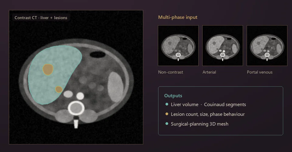
Illustration: abdominal CT slice with the liver outlined in teal and two lesions in gold, beside three contrast-phase thumbnails and a list of outputs.

Organs & anatomyStarter

LiverScope: liver and lesions across contrast phases

Segment the liver, its vessels and every focal lesion from multi-phase CT. Then turn the masks into numbers surgeons use: Couinaud segments, lesion size and the volume of liver left after resection.

You will learn 3D U-Net and nnU-Net, multi-phase fusion, lesion-wise evaluation.

CTLiTS3D segmentation
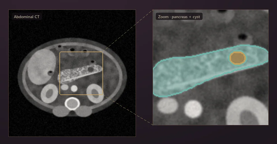
Illustration: abdominal CT with a zoomed view of the pancreas in teal and a small cyst in gold.

Organs & anatomyIntermediate

PancreasFinder: a small organ, an early signal

The pancreas is small, thin and shaped differently in every patient. Find it with a coarse-to-fine model, segment it, and flag small cystic lesions early, when they matter most.

You will learn cascaded localisation, small-object segmentation, class imbalance.

CTMRIMSD Pancreas
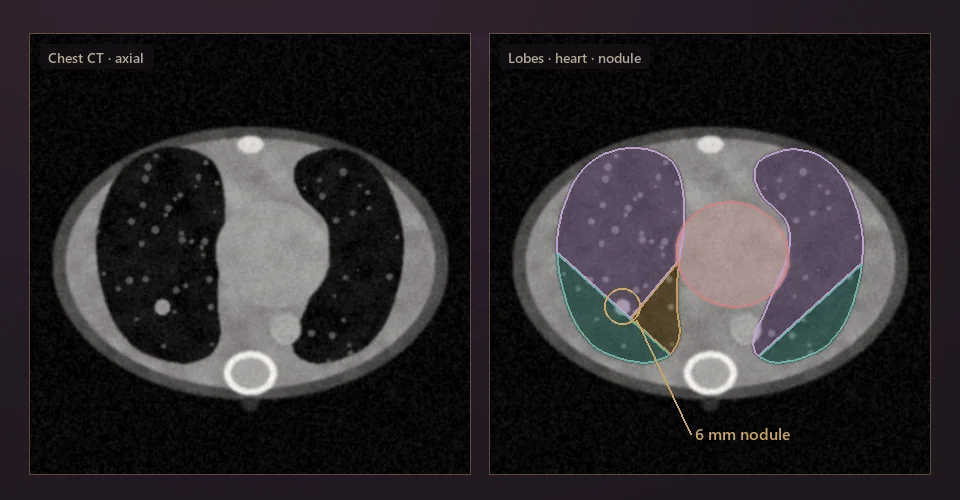
Illustration: axial chest CT, raw and with lung lobes, heart and a small nodule highlighted.

Organs & anatomyIntermediate

ThoraxNet: one pass through the chest

Lobes, airways, heart and nodules from a single chest CT, with a chest X-ray branch for settings without CT. Airways test whether a model respects thin, connected structures.

You will learn multi-structure segmentation, topology-aware losses, nodule detection.

CTX-rayLIDC-IDRI
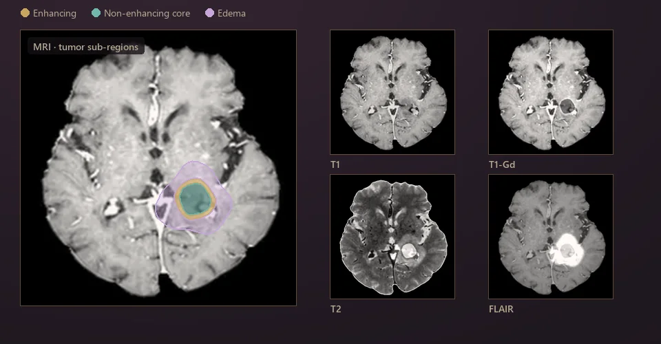
Illustration: brain MRI with tumor sub-regions (enhancing, core, edema) and four MRI sequences: T1, T1-Gd, T2 and FLAIR.

Organs & anatomyIntermediate new

PedsBrain: tumor segmentation built for children

Brain tumors in children look and behave differently from adult ones, and far less data exists. Transfer what adult models know, then adapt them to the pediatric sub-regions that guide treatment.

You will learn multi-sequence MRI, transfer learning, learning from small datasets.

MRIBraTS-PEDsTransfer learning
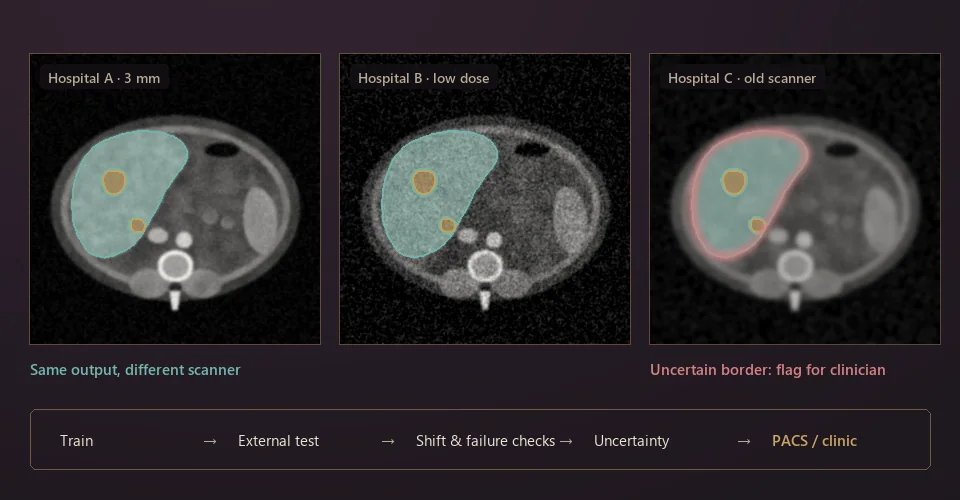
Illustration: the same liver segmentation on scans from three hospitals, one with an uncertain border flagged for review, above a pipeline from training to the clinic.

Clinical segmentationIntermediate

Bench to Bedside: segmentation that survives the next hospital

A model that scores well on one dataset often fails on another scanner. Test organ and tissue models across sites, detect the shift, estimate uncertainty and flag cases for a clinician instead of failing silently.

You will learn external validation, domain generalisation, calibration and uncertainty.

Multi-siteUncertaintyDeployment
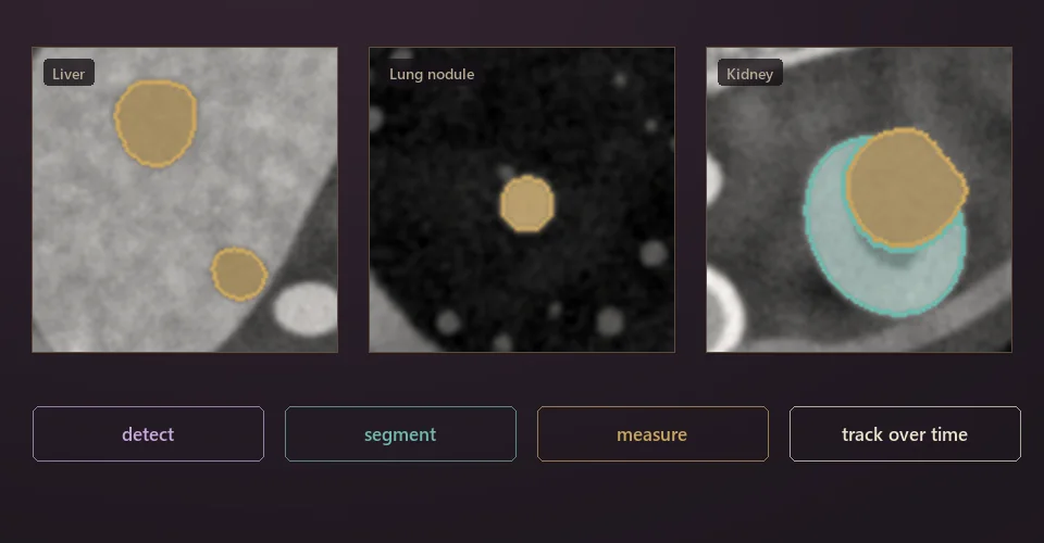
Illustration: zoomed tumors in the liver, lung and kidney, with the steps detect, segment, measure and track over time.

Clinical segmentationIntermediate

Every Lesion Counts: tumor segmentation across organs

Small, faint tumors are the ones models miss and clinicians care about. Detect, segment and measure lesions in liver, lung and kidney, and follow them from one scan to the next.

You will learn detection plus segmentation, lesion-level metrics, longitudinal registration.

CTKiTSLiTS
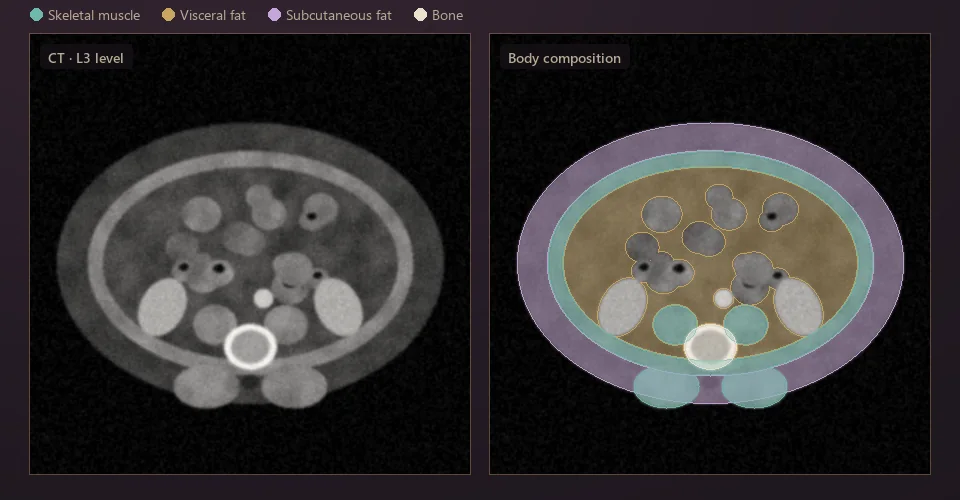
Illustration: CT slice at the L3 vertebra with skeletal muscle, visceral fat, subcutaneous fat and bone segmented.

Clinical segmentationStarter

BodyComp: muscle and fat from routine scans

Every abdominal CT already contains information on muscle and fat. Segment it automatically to screen for sarcopenia and metabolic risk, using scans that were taken for another reason.

You will learn tissue segmentation, slice selection, linking imaging biomarkers to outcomes.

CTOpportunistic screening
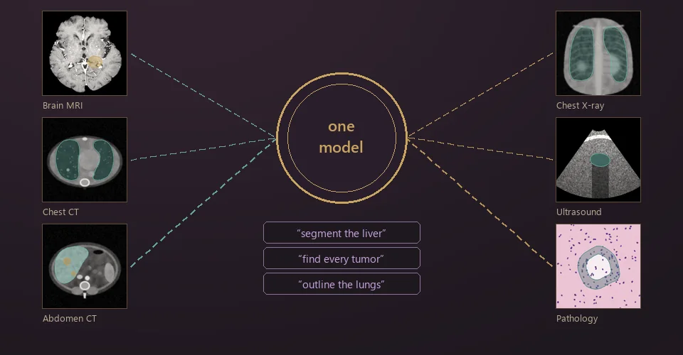
Illustration: brain MRI, chest CT, abdominal CT, chest X-ray, ultrasound and pathology images all feeding one model, which takes text prompts.

Foundation modelsAdvanced

OmniSeg: one promptable model for every organ

One model for CT, MRI, X-ray, ultrasound and pathology, prompted with a click or a sentence such as “segment the liver”. Compare it honestly with SAM, MedSAM and specialist models.

You will learn foundation models, prompting, training on many datasets at once.

Multi-modalSAM / MedSAMText prompts
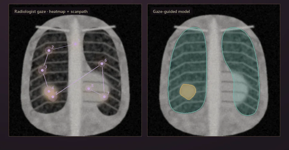
Illustration: chest X-ray with a radiologist's gaze heatmap and numbered scanpath, beside a model's lung and lesion segmentation guided by that gaze.

Human signalsIntermediate

GazeGuide: learning from where radiologists look

Eye trackers record where a radiologist looks while reading. Use that gaze as free supervision to teach models where to attend, and predict gaze to learn how experts search an image.

You will learn eye-tracking data, attention supervision, human–AI collaboration.

Eye trackingX-rayREFLACX
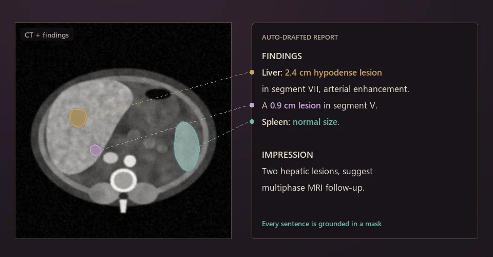
Illustration: CT slice with findings linked by dashed lines to sentences in an automatically drafted radiology report.

LanguageAdvanced rising

GroundedReport: reports a radiologist would sign

Draft realistic radiology reports in which every finding points to a mask or a measurement. Catch hallucinated findings, and evaluate with clinical metrics rather than word overlap.

You will learn vision-language models, grounding, clinical evaluation of text.

LanguageCTMIMIC-CXR
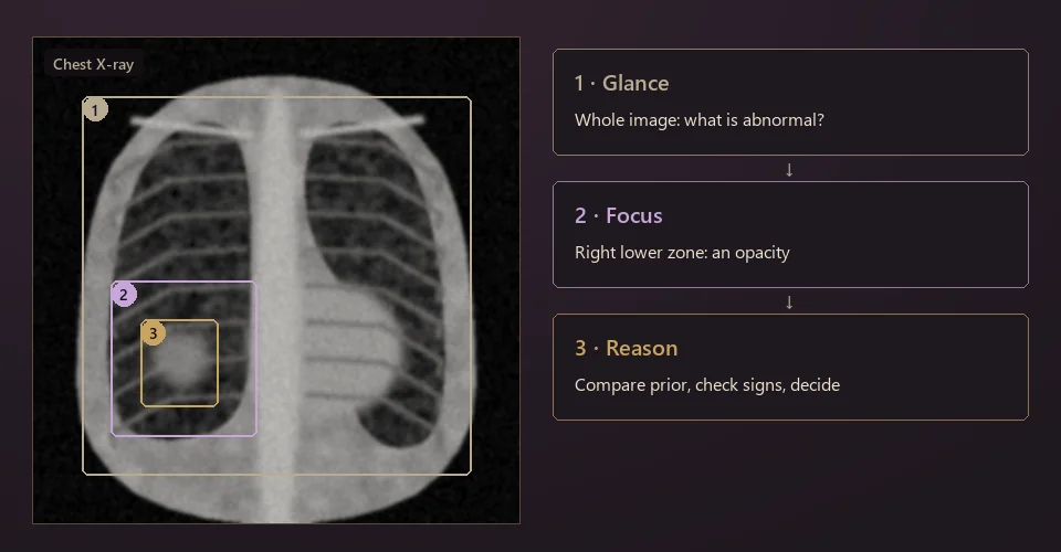
Illustration: chest X-ray with three nested boxes for glance, focus and reason, beside three steps describing an expert's reading process.

Cognitive AIAdvanced frontier

Think Like a Radiologist: search-then-reason agents

Experts glance at the whole image, focus on what looks wrong, then reason with priors and history. Build agents that read images in the same order and can explain each step.

You will learn agentic vision-language models, visual reasoning, ideas from cognitive science.

CognitionAgentsReasoning
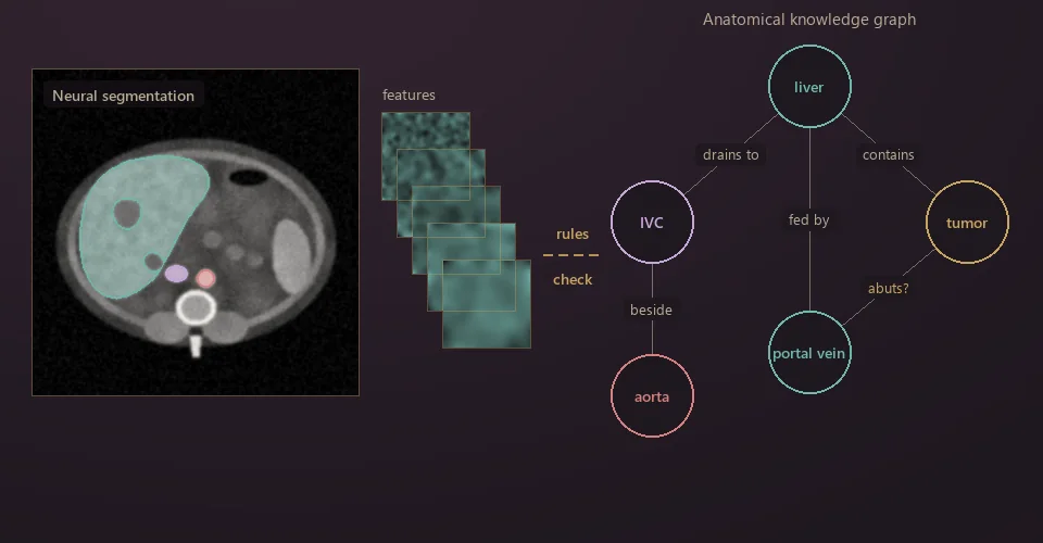
Illustration: a neural segmentation of abdominal CT and its feature maps, checked against a knowledge graph of anatomical relations such as liver, IVC, aorta, portal vein and tumor.

Hybrid AIAdvanced frontier

Anatomy-Aware Hybrid AI

Neural networks sometimes produce anatomy that cannot exist. Pair them with a knowledge graph of anatomical rules (what touches what, what contains what) to catch those errors and explain decisions.

You will learn neuro-symbolic methods, knowledge graphs, constraint-based refinement.

KnowledgeCTNeuro-symbolic

The images are synthetic illustrations made from simple shapes and noise, not patient data. The brain figure uses a public MRI slice. Datasets named on the cards are examples to start from.

Past cohort: AIO 2025 Mentorship Program (6 projects, applications closed)

Open to AIO 2025 students only (deadline January 28, 2026; interviews January 29 – February 1).

Project 1 · 5–6 members

Segment Anything Model under Noisy Medical Radiology Imaging Conditions

Robustness, stability and failure modes of SAM, MedSAM and SAM2 on 2D medical images under controlled noise. Target: a CVPR workshop benchmark, then a Q1 journal.

Project description
Project 2 · 5–6 members

Segment Anything Model (SAM) under Occluded Medical Imaging Conditions

An occlusion-robustness benchmark for SAM-based models, using surgical tools, probes and implants as occluders. Target: a CVPR workshop, then a Q1 journal.

Project description
Project 3 · 4 members

Brain Image Segmentation under Annotation Scarcity


How brain segmentation degrades as labeled data shrinks, across classical and advanced architectures. Target: an international conference, then a Q2 journal.
Project description

Project 4 · 4 members

3D Liver Image Segmentation under Limited Annotation


Liver and liver-tumor CT segmentation on LiTS with progressively fewer labels. Target: an international conference, then a Q2 journal.

Project description
Project 5 · 4 members

Semi-Supervised Breast Cancer Ultrasound Classification

CNNs versus Vision Transformers for breast-ultrasound classification under limited labels. Target: an international conference, then a Q2 journal.

Project description
Project 6 · 4 members

Few-Shot White Blood Cell Image Classification

White-blood-cell classification from blood-smear images with few labels, CNN versus ViT. Target: an international conference, then a Q2 journal.

Project description

V

Our People

Mentors lead our research groups and run the community day to day: PhD students, postdocs and faculty at universities and medical centers in the United States and Australia. Each appears here as an animal alter ego, drawn for this page.

Mentors

Cartoon owl
Phat K. Huynh, PhD

Assistant Professor, North Carolina A&T State University

Cartoon fox
Hien Kha, MD-PhD

Postdoc, MD Anderson Cancer Center

Cartoon deer
Minh Le, MD-PhD

Postdoc, Yale University

Cartoon bear
Minh Gia Hoang, PhD

Postdoc, Mayo Clinic

Cartoon rabbit
Luu Le

Research Fellow, Northwestern University

Cartoon corgi
Thien H. Nguyen

Research Associate, Carnegie Mellon University

Cartoon raccoon
Thinh B. Lam

PhD Student, UNC Charlotte

Cartoon white cat
Vi Vu

PhD Student, Duke University

Cartoon hedgehog
Khai Bui

PhD Student, Duke University

Cartoon otter
Dat Chung

PhD Student, Florida International University

Cartoon red panda
Phuc Nguyen

PhD Student, UNC Chapel Hill

Cartoon koala
Quan Nguyen

PhD Student, University of Technology Sydney

Last but not least, thank you to every mentee who has worked alongside us and contributed so much to this community. We could not have come this far without you.

VI

Our Network

Where the people behind AIMA work: our mentors, and the advisors, professors and collaborators who support our groups and are not listed by name. Almost everyone started in Vietnam. From three hubs there, the community now reaches 33 institutions in 8 countries on 4 continents.

  • 3hubs in Vietnam
  • 33institutions
  • 29cities
  • 8countries
  • 4continents

Loading the map…

Hanoi, Da Nang and Ho Chi Minh City, where most of us set out from, linked to every city in the network. Hover or tap a dot to see the institutions there. Which hub a city links to is illustrative.

Northwestern University Harvard University Carnegie Mellon University Duke University Yale University Johns Hopkins University University of Pennsylvania Mayo Clinic
University of Chicago Cornell University The University of Texas at Austin MD Anderson Cancer Center University of North Carolina at Chapel Hill University of Pittsburgh University of Wisconsin–Madison Stony Brook University
University of North Carolina at Charlotte University of Houston University of Alabama at Birmingham University of Central Florida Florida International University North Carolina A&T State University Université Bourgogne Europe Université Claude Bernard Lyon 1 Aalto University
Nanyang Technological University Taipei Medical University National Taiwan University National Cheng Kung University Sejong University Mohamed bin Zayed University of Artificial Intelligence University of Technology Sydney Swinburne University of Technology
United States · 22Northwestern · Harvard · Carnegie Mellon · Yale · Duke · Johns Hopkins · Penn · Cornell · Chicago · Mayo Clinic · MD Anderson · Pittsburgh · Wisconsin–Madison · Stony Brook · UT Austin · UNC Chapel Hill · UNC Charlotte · NC A&T · UAB · Houston · UCF · FIU
Europe · 3Université Bourgogne Europe (Dijon) · Université Claude Bernard Lyon 1 · Aalto University (Finland)
Middle East · 1MBZUAI (Abu Dhabi)
Asia · 5Nanyang Technological University (Singapore) · Taipei Medical University · National Taiwan University · National Cheng Kung University (Tainan) · Sejong University (Seoul)
Australia · 2University of Technology Sydney · Swinburne University of Technology (Melbourne)

VII

Contact

Want to join the next cohort? It will be announced here and on our Facebook page. If one of the projects above excites you, write to us with the project name, a short note on your background, and what you want to learn.

AIMA Research
Cohorts, projects & student enquiries
Facebook
Announcements & openings